We appreciate the value in combining art with science to reach a broader audience. Below are images from the Dewey Lab which connect these two principles. Keep checking back for new content captured and created by current researchers!
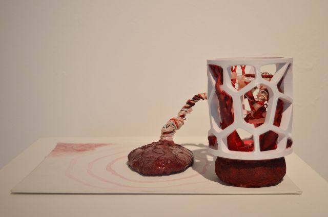
Placenta Power- People's Choice Award Winner for the UCSB 12th Annual Art of Science competition (Andrea Gomez, 2025)
Andrea’s sculpture is inspired by research using placentas as a biomaterial to aid with bone regeneration. Inflammation and infection hinder the wound healing process, nevertheless Dr. Dewey and her team found that there are compounds in the outermost layer of the placenta, the amnion, that fight chronic inflammation and promote healthy bone cell growth. These compounds include collagen, calcium, phosphates, and other bioactive factors. Then, they are combined into a jelly-like mineralized collagen scaffold that is held in place by a microscopic 3D printed structure (made with PLA filament). The placenta has the universal potential to help with essential wound healing, and to power innovative, surreal advancements in biomedical research. The making of this project paralleled the framework of the scientific process and similarly involved mindful, hands-on research. As a scientist and artist, Andrea aims to embrace the beautiful, entangled dynamic between STEM research and the creative process within her work.
Life Imitates Art: Coral Cells On a Sea of Collagen- Image submitted to the UCSB 12th Annual Art of Science competition (Renata Dos Reis Marques, 2025)
False-colored scanning electron microscopy image of coral fragment-derived cells cultured on a bovine collagen I hydrogel scaffold.

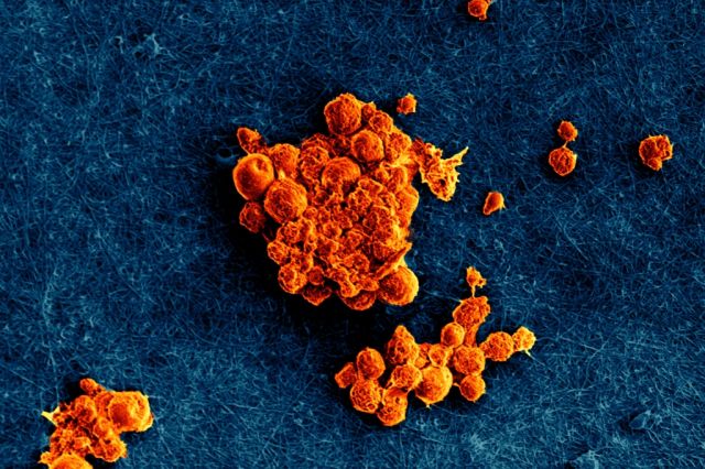
Human fibroblasts secreting matrix-bound nanovesicles- Image featured on the cover of the Journal of Extracellular Biology (Dr. Dewey, 2024)
False-colored scanning electron microscopy image of human fibroblasts secreting matrix-bound nanovesicles (MBV, orange), a type of extracellular vesicle embedded in the extracellular matrix.
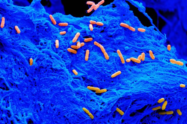
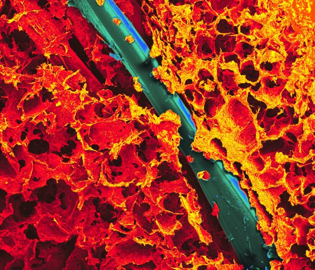
Bacteria cells on a collagen scaffold- Image Featured on National Science Foundation Multimedia Gallery (Dr. Dewey, 2023, UIUC)
Cells of Pseudomonas aeruginosa (orange), a harmful species of bacteria, are scattered over the surface of a collagen scaffold (blue).

Hyperelastic Bone 3D-print- Society for Biomaterials ECTM SIG Sci-Art Image Competition Winner (Dr. Dewey, 2022, UIUC)
This is a false-colored SEM image of a 3D-print fabricated by Dimension Inx LLC (Chicago, IL). This material has been used for bone repair applications as the mineral crystals and unique polymer chemistry allow for flexibility and porosity.
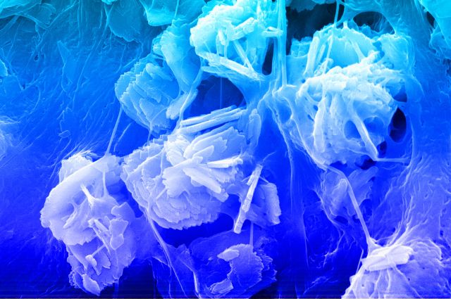
Mineralized collagen scaffold showing mineral crystals and collagen fibers- Image Featured on National Science Foundation Multimedia Gallery (Dr. Dewey, 2022. UIUC)
Electron microscope (SEM) image of a mineralized collagen scaffold showing mineral crystals and collagen fibers. The color was added by researchers.

Hyperelastic Bone Galaxy- Beckman Institute Graduate Student Image of Research Contest Winner (Dr. Dewey, 2020, UIUC)
This is a false-colored SEM image of a 3D-print fabricated by Dimension Inx LLC (Chicago, IL). This material has been used for bone repair applications as the mineral crystals and unique polymer chemistry allow for flexibility and porosity.
Mineralized collagen-PLA composite- Finalist for the Graduate Image of Research Contest (Dr. Dewey, 2018, UIUC)
Mineralized collagen scaffolds are soft, porous materials used for bone repair, but are mechanically weak. To better match mechanical properties of bone, a polylactic acid (PLA) 3D-print was embedded within the collagen to create a composite with stiffer properties while still allowing for cell infiltration. This is an SEM image demonstrating the 3D-print (blue) within the collagen (orange).